
Smart phones seriously harm the developing brains of children and adolescents: time to take action
Thu 2:05 pm +00:00, 19 Nov 2026The interview below was conducted in German and was triggered by new lab findings and a 2024 animal study. It first appeared in the diagnose:funk Kompakt magazine (Issue 1/2025) and online on 2 March 2025. The questions were asked by Peter Hensinger, Executive Director of diagnose:funk, an environmental and consumer protection organisation based in Germany.
The goal of diagnose:funk is to educate about the health- and environmental-damaging effects of electromagnetic fields, such as those caused by mobile phones, smartphones, mobile phone antennas, WLAN, DECT and other electrosmog sources, as well as the psycho-social effects of digital media. This is intended to change consumer and political behaviours and promote solutions for environmentally friendly and future-proof technologies.
Recommendations: The researcher urges schools, parents, and caregivers to:
– Cut screen time sharply
– Favour hands-on and social learning over digital
– Use wired (not Wi-Fi) alternatives where possible
– Keep wireless devices away from bedrooms
– Make sure children understand the risks
– She argues the precautionary principle — “if there’s a plausible risk, protect the vulnerable” — should apply here.

Photo by bruce mars on Unsplash
“It is high time that the negative effects of high-frequency EMFs on the brain development of children and adolescents are taken seriously!” – Interview with neurobiologist Dr. Keren Grafen
Note: significant findings explained in the article have been highlighted in italics throughout
In her lecture in Neckartenzlingen, neurobiologist Prof. G. Teuchert-Noodt explained that brain metabolism is largely controlled by electromagnetic frequencies between 4 Hz and 30 Hz and that homeostasis in the brain depends on a delicate interplay of these frequencies. Her institute has published research findings on this topic. It therefore follows logically that external radiation, such as that generated by mobile phone radiation, would affect this process.

Dr. Keren Grafen was a researcher at Prof. G. Teuchert-Noodt’s institute and is very familiar with the neurobiological research conducted there. Peter Hensinger interviewed her about the current state of research on the effects of mobile phone radiation (EMF) on brain metabolism.
Dr. Keren Grafen, speaking in her personal capacity
Neurobiology has demonstrated how sensory overload from digital media influences brain metabolism, inhibits the development of the prefrontal cortex, and can even lead to addiction. Prof. Gertraud Teuchert-Noodt explains this in her video lecture. The study by Kim et al. (2024), “High-Frequency Exposure Induces Synaptic Dysfunction in Cortical Neurons, Causing Changes in Learning and Memory in Early Postnatal Mice,” now also demonstrates, at the molecular level, the pathological effects of high-frequency radiation on brain development in the prefrontal cortex (frontal lobe). Mobile phone radiation inhibits the development of synaptic structure and density as well as neurite growth, with negative consequences for behaviour, spatial learning, and memory.
Negative effects of mobile phone radiation on memory have been demonstrated not only in animal studies. A study titled “A Prospective Cohort Study on Memory Performance in Adolescents and the Individual Brain Dose from Microwave Fields Due to Wireless Communication,” involving 700 adolescents in Switzerland, found that high-frequency electromagnetic fields from mobile phones have an adverse effect on the development of memory performance in both figural and verbal memory. It was conducted by the Swiss Tropical and Public Health Institute (Förster et al. 2018, Schoeni et al. 2015). Mobile phone use among 12- to 17-year-olds was evaluated over the course of one year. As expected, frequent callers exhibited increased radiation exposure to the brain. The intriguing finding: the more phone calls made, the poorer the performance on the figural memory test.
Verbal memory also showed poorer results.
Have Kim et al. now provided a neurobiological explanation for the findings of Förster et al.? What is the significance of these study results? We asked neurobiologist Dr. Keren Grafen about this.
The Hippocampus—The Center for Effective Learning
DIAGNOSE:FUNK: Dr. Grafen, we’d like to ask you to assess the results of the two studies by Kim and Förster, both of which investigated the effects of mobile phone radiation on the brain. First, for our readers who aren’t biologists, what is the function of the hippocampus? What role do synapses and neurites play in the brain?
KEREN GRAFEN: I’d be happy to! The hippocampus is a fascinating structure in the brain that plays a crucial role in short-term memory, the transfer of information to long-term memory, as well as in emotions, motivation, and spatial orientation. The name “hippocampus” derives from its shape, which resembles a seahorse.
A remarkable feature of the hippocampus is its ability to generate new nerve cells throughout a person’s lifetime. This occurs in an embryonic germ layer located within the hippocampus, which enables continuous neurogenesis. This process contributes significantly to neural plasticity by maintaining the adaptability of the neural network and preventing the formation of rigid structures.
Since the hippocampus must continuously store new information, the system is necessarily receptive to environmental stimuli. This phenomenon, known as hippocampal neurogenesis, is a central area of research that I have studied intensively for many years. It is important to note that the formation of new neurons in the hippocampus continues well into late adulthood, serving as an essential prerequisite for learning processes, emotional regulation, and cognitive flexibility.
Another key function of the hippocampus is its role in the creation of cognitive maps. The discovery of place cells in the hippocampus and grid cells in the adjacent entorhinal cortex was honored with the 2014 Nobel Prize in Physiology or Medicine. These specialized neurons are essential for encoding spatial information and enable the calculation of internal maps for navigation.
The neuroanatomical basis for all these processes is formed by neurites—that is, axons and dendrites—which ensure extensive connectivity between neurons. Synapses play a crucial role in signal transmission and enable the exchange of information within neural networks.
Damage to the hippocampus has far-reaching consequences for cognitive and spatial processes. Experimental studies in rodents show that without this structure, effective learning is no longer possible – a finding that has also been confirmed in humans. The case of patient H.M., who underwent bilateral hippocampal resection in the 1950s, vividly illustrates the central importance of this region: After the procedure, he was unable to form new memories or orient himself spatially.
RF-EMF Causes the Branches of Neural Trees to Atrophy
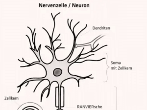
Graphic:diagnose:funk
DIAGNOSE:FUNK: A finding from the study by Kim et al.: RF-EMF exposure led to a reduction in neurite length and the number of branches. What consequences might this have?
KEREN GRAFEN: A neural network can be likened to a forest: Each nerve cell resembles a tree whose branches extend far and wide and connect with other trees. However, when individual branches—or even entire trees—die off, gaps appear in the dense forest—the once-vibrant connection between the trees is severed. The study by Kim et al. was able to impressively demonstrate precisely this phenomenon: Exposure to radiofrequency electromagnetic fields (RF-EMF) has a negative effect on the axons and dendrites of nerve cells in the hippocampus. The branches of the neural trees atrophy, their number decreases, and the network loses stability.
Even more profound are the potential effects on spatial orientation: EMF exposure can impair both the formation and the function of cognitive maps. This would have implications not only for our memory but also for more complex thought processes—such as the ability to distinguish between the past and the future, or social interaction with other people. Just as a damaged forest not only alters its ecosystem but also affects the lives of countless animals, disrupted neural connectivity could have far-reaching consequences for our thinking and behaviour.
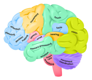
Graphic:Queensland Brain Institute https://qbi.uq.edu.au/brain/brain-anatomy/lobes-brain
Hebb’s learning synapse: “Neurons that fire together, wire together.”
DIAGNOSE:FUNK: The expression of AMPA and NMDA glutamate receptors (Hebb’s learning synapse) was significantly reduced in the hippocampal neurons studied. What does this mean? Can the implications for neuroplasticity also be inferred from this? What role does neuroplasticity play?
KEREN GRAFEN: To answer that, we should first explain the Hebb learning synapse, which represents a fundamental principle of neural plasticity and was formulated by the Canadian researcher Donald Hebb in 1949: “Neurons that fire together, wire together.” This means that the synaptic connection between two neurons is strengthened when they are repeatedly active at the same time. More specifically: When a presynaptic neuron (sender) is active at the same time as a postsynaptic neuron (receiver), the synaptic connection is strengthened. This phenomenon is known as long-term potentiation (LTP). The NMDA receptor plays a central role in this process.
The process unfolds as follows: An incoming signal first activates the AMPA receptor, which immediately transmits the excitation. Only when the neuron remains active over an extended period—as is the case during learning processes involving repeated stimulation—is the NMDA receptor activated. A distinctive feature of the NMDA receptor is that it is blocked by a magnesium ion in its resting state. Only when the input to the receiving neuron is strong enough is this block lifted. Through various chemical processes, this leads to structural changes at the synapse: it grows, becomes more stable, and increases in size. These changes facilitate signal transmission at this synapse, thereby increasing the efficiency of learning. This is what Hebb meant in 1949 when he postulated that the synaptic connection between two neurons is strengthened when they are repeatedly active simultaneously.
The significantly reduced expression of AMPA and NMDA glutamate receptors in hippocampal neurons—as Kim demonstrated in the study mentioned above—means that learning processes are impaired at the physiological level. This, in turn, results in the brain’s ability to adapt structurally and functionally to experiences and environmental factors—a process known as neuroplasticity—being severely limited. The consequences are far-reaching: the anatomical basis for all learning is impaired.
HF-EMFs influence the homeostasis of brain rhythms
DIAGNOSE:FUNK: At the Teuchert-Noodt Institute, where you worked, a study by Hoffmann et al. in 2001 discovered an electrophysiological correlation: Brain metabolism is regulated by EMFs in the frequency range of 4 Hz to 30 Hz. Could you explain this mechanism to us, and could the damage discovered by Kim et al.—including reduced BDNF expression—also be related to this?
KEREN GRAFEN: The electrophysiological correlation discovered by Hoffmann et al. (2001) at the Teuchert-Noodt Institute shows that electromagnetic fields (EMFs) in the frequency range of 4 Hz to 30 Hz influence hippocampal neurogenesis. It is particularly striking that EMF exposure in the frequency range of 1, 29, and 50 Hz significantly reduces neurogenesis, while other frequencies, such as 8 and 12 Hz, have no effect whatsoever. The study interprets this to mean that only certain frequencies activate the release of neurotransmitters and hormones, which in turn control the changes in the hippocampus.
This finding paves the way for an interesting hypothesis: There is a common mechanism found in both the study by Hoffmann et al. and the study by Kim et al.: EMF-mediated regulation of neurotransmitters and hormones could be the trigger for reduced BDNF expression and, consequently, decreased synaptic density—an extremely interesting hypothesis that will certainly be the focus of future research. BDNF, or Brain-Derived Neurotrophic Factor, is a protein that plays a central role in the formation of new synapses in the brain. A deficiency in BDNF is associated with cognitive deficits and neurodegenerative diseases.
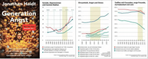
Jonathan Haidt: Tipping Point 2012: Smartphone Use and Its Catastrophic Effects on Children and Adolescents
Graphic 1: Suicide, depressiion and social media
Graphic 2: Loneliness, fear and stress
Graphic 3: Getting together with friends, close friends, self-satisfaction
Graphics: diagnose:funk
The brain learns primarily through movement, through touching and grasping—and in three dimensions.
DIAGNOSE:FUNK: What role does all of this play in learning and healthy brain development? Based on your neurobiological knowledge, what do you think when you see children and adolescents constantly on their phones?
KEREN GRAFEN: That’s a very important question: From a neurobiological perspective, healthy brain development plays a central role in learning, because the brains of children and adolescents are particularly plastic and malleable. This means that, on the one hand, they are especially receptive, but on the other hand, they are also particularly vulnerable to harmful influences. To use the metaphor of a forest, every new experience, every learning opportunity, and every interaction is integrated into this neural network as new “branches.” Harmful or inappropriate stimuli cause this forest to wither away. It is not only harmful influences—such as the damaging effects of EMF radiation described above—that play a role here, but also the way in which learning takes place. The brain learns primarily through movement, through touching and grasping—and it does so in three dimensions. This means that we do not merely passively absorb information, but rather integrate it and anchor it in our brains through active, physical engagement with the environment.
The growing dependence on digital devices and the associated 24/7 EMF exposure could thus negatively impact neural growth and cognitive abilities, particularly in the highly vulnerable brains of our children and adolescents. In neurobiology, we refer to such cases as “premature maturation,” as seen, for example, in the Kasper Hauser effect. Therefore, from a neurobiological perspective, it is high time to take these experimental studies seriously, reduce EMF exposure, and utilize alternative methods that support learning and brain development by promoting movement and interaction with the real world.
DIAGNOSE:FUNK: All of this implies that brain development would be severely damaged by mobile phone radiation. The study was conducted on mice. Can we draw conclusions from this regarding a risk to our children’s brain development?
KEREN GRAFEN: Yes, I would. Although the studies cited above were conducted on mice, the fundamental neurobiological mechanisms—particularly those involving the hippocampus, a key relay station—including neurogenesis, synaptic plasticity, and spatial coordination—are entirely comparable in mammals, including humans. If these findings are ignored by a political establishment tainted by industry interests, they should at least serve as a warning signal for parents, teachers, and educators. It is high time that the negative effects of high-frequency EMFs on the brain development of children and adolescents are finally taken seriously. Precautionary measures such as reducing mobile phone use among children, using wired alternatives, and minimizing radiation exposure in the sleeping area are the bare minimum required to prevent potential long-term damage.
________________________________________________________________________
In the video, Prof. Dr. Michael Kundi explains the significance of the findings from the Swiss study by Foerster et al. on the impairment of figural thinking, at the 26:00 mark: https://youtu.be/07G65fE0xEM?si=IZb9t7iyde2_p3Qh

Swiss Study: Smartphone Use Impairs Figurative Memory
DIAGNOSE:FUNK: Kim et al. have researched biological processes in mice. This could retrospectively explain the results of the study by Förster et al. (2018) from Switzerland. The study examined the effect of RF-EMF on memory performance in approximately 670 adolescents. The main findings: An increase in cumulative RF-EMF exposure was associated with a decline in figural memory performance. The effect was particularly pronounced among participants who preferred to use their mobile phones on the right side of their heads. The effect was significant when RF-EMF doses were estimated based on carrier data.
KEREN GRAFEN: Yes, figural memory refers to the ability to store and recall visual impressions such as images, shapes, patterns, or spatial structures. It is an essential part of visual memory and allows us to retain the appearance of objects, faces, places, and situations without relying on verbal descriptions. It plays a key role in spatial orientation and the reconstruction of perceptions. And this is precisely where the hippocampus comes into play—THE central hub for spatial processing and orientation; do you remember place cells? A clear connection can therefore be established.
The study by Kim et al. is therefore consistent with the findings of Förster et al. (2018), who uncovered a link between cumulative RF-EMF exposure and a decline in figural memory performance among adolescents. Over the long term, changes in the hippocampus disrupt the complex three-dimensional neural network.
At this point, however, I would like to make a critical remark: The hypothesis that certain brain regions—such as the right hippocampus—are responsible for specific functions like figural memory is a simplified view. Personally, I consider this “left-right hypothesis” problematic, as we should increasingly shift from a strictly anatomical perspective toward a systemic and networked way of thinking about the brain. The brain does not function in isolation within individual areas, but rather in dynamic, interactive networks. This perspective shows us that the effects of EMFs are much more complex and go far beyond the isolated consideration of brain regions.
________________________________________________________________________
Lecture by Prof. Leif Salford on his studies regarding the effects of mobile phone radiation on the brain (in English): https://youtu.be/E_WJ_aJPWIA?si=V9Felg9HPxcYCtaD
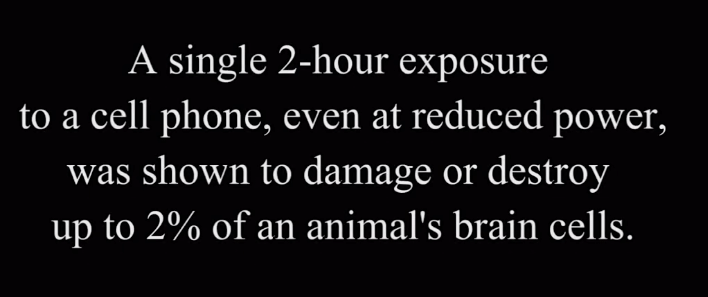
Salford studies confirm: RF-EMFs open the blood-brain barrier
DIAGNOSE:FUNK: In 2022, you yourself published the article “Albumin as a Key Marker: How the Permeability of the Blood-Brain Barrier Changes Following Exposure to Cell Phone Radiation” with Thieme, in which you write: “Professor Salford’s findings on albumin extravasation caused by electromagnetic fields may provide insight into pathogenic mechanisms for a broad spectrum of neurological disorders associated with blood-brain barrier dysfunction.” When viewed in conjunction with the findings of Kim et al., what role might this mechanism play in brain development, especially when even young children are using smartphones?
KEREN GRAFEN: Yes, the extremely insightful findings of Prof. Leif Salford—which I included in my article “Albumin as a Key Marker” and combined with current data—highlight the immensely complex and potentially dangerous effects of EMFs on the brain—and in a completely new way: via the blood-brain barrier. It is now well documented that the extravasation (leakage) of albumin is associated with hyperpermeability of the blood-brain barrier (BBB).
When these findings are combined with the results of the previously mentioned studies, the seriousness of the potential effects of EMFs on children’s brain development becomes even clearer. The hyperpermeability of the blood-brain barrier caused by EMFs opens the brain to harmful substances that place an additional burden on the brain, particularly at the immunological level (Note d:f: the new study by Kizilçay et al. (2025) once again confirms this effect of EMFs). The neural network, which is still developing and particularly vulnerable, is thereby further and severely disrupted.

Photo by Lance Anderson on Unsplash
And now, please imagine what will become of our children’s future if ever-younger children are exposed to smartphones and other wireless devices! The mechanism of BHS dysfunction, combined with the findings of Kim et al., shows that chronic EMF exposure leads to significant impairments in brain function. I see this every day in my practice: desperate parents watching helplessly as their children act like remote-controlled beings. It is high time that at least parents, teachers, and educators become aware of the ever-growing danger that EMF exposure poses to the brains of our children and adolescents. There are alternatives, even in an increasingly digital world. Switching to wired devices, drastically reducing mobile phone use—especially among young children—and, above all, providing urgently needed education about the risks of EMFs are essential to protecting healthy brain development. We can no longer stand idly by while our children’s health is put at risk!
It is high time to address the dramatic consequences
DIAGNOSE:FUNK: In his book *Generation Angst*, Jonathan Haidt argues that since 2012, smartphone use has caused young people’s mental health to plummet. The PISA studies show that skills in math, writing, reading, and speaking are in free fall, while mental health issues among children and adolescents are on the rise. Doesn’t the study by Kim et al. provide a plausible explanation for this? What consequences do you see arising from these findings for daycare centres, schools, and parents?
KEREN GRAFEN: Yes, the study by Kim et al. not only provides a plausible biological explanation for the arguments put forward by Jonathan Haidt in *Generation Angst*, but also allows us to recognize the full extent of the effects of EMF on the mental health of young people. Haidt notes that the increasingly frequent use of smartphones since 2012 has led to a dramatic decline in adolescents’ mental health, as reflected in rising rates of mental illness and declining academic performance. The numerous neurobiological changes I described above could be the cause of adolescents’ growing difficulties in basic cognitive areas such as arithmetic, reading, writing, and speaking.
At the neuroanatomical level, the hippocampus, in conjunction with the amygdala, plays a crucial role in the perception of fear. Higher-order structures such as the prefrontal cortex can help contextualize fear, allowing us to assess whether it is real or imaginary. As explained in Prof. Dr. Teuchert-Noodt’s lecture in Neckartenzlingen, the mesolimbocortical dopaminergic stress pathway plays a crucial role in this process. This stress pathway determines how we cope with stress and anxiety, and its impairment dramatically reduces the emotional resilience of young people.
It is high time that we address the dramatic consequences of these findings! Schools, daycare centres, and parents must act now to reduce the unchecked EMF exposure of children and adolescents. Urgent measures are needed to protect the well-being of our children: We must drastically reduce screen time, promote analog learning methods and social interactions, raise awareness, and foster healthier use of digital devices. If we fail to act, we risk causing permanent damage to the mental health and cognitive development of the next generation.
DIAGNOSE:FUNK: Dr. Grafen, thank you very much for explaining these highly controversial connections. These findings from their research need to be made known. We will share this interview with as many parents, educators, and healthcare professionals as possible.
References
Hoffmann K, Bagorda F, Stevenson AF, Teuchert-Noodt G (2001): Electromagnetic exposure affects hippocampal dentate cell proliferation in gerbils (Meriones unguiculatus), Indian J Exp Biol 39(12): 1220-1226.
Foerster M, Thielens A, Joseph W, Eeftens M, and Röösli M (2018): A Prospective Cohort Study of Adolescents’ Memory Performance and Individual Brain Dose of Microwave Radiation from Wireless Communication. Environmental Health Perspectives, Vol. 126, No. 7, Research Open Access, https://www.emf-portal.org/de/article/35641
Kim JH, Seok JY, Kim YH, Kim HJ, Lee JK, Kim HR (2024): Exposure to Radiofrequency Induces Synaptic Dysfunction in Cortical Neurons, Causing Alterations in Learning and Memory in Early Postnatal Mice. International Journal of Molecular Sciences, 25(16). https://www.mdpi.com/1422-0067/25/16/8589
Lehmann K, Grund T, Bagorda A, Bagorda F, Grafen K, Winter Y, Teuchert-Noodt G (2009): Developmental effects on dopamine projections and hippocampal cell proliferation in the rodent model of postweaning social and physical deprivation can be triggered by brief changes in environmental context. Behav Brain Res 205(1): 26–31.
Neufeld J, Teuchert-Noodt G, Grafen K, Winter Y, Witte AV (2009): Synaptic plasticity in motor, sensory, and limbic-prefrontal cortex areas as measured by degrading axon terminals in an environmental model of gerbils (Meriones unguiculatus). Neural Plast 2009: 1-15.
Teuchert-Noodt G, Hensinger P (2025): There is no way out of the smartphone epidemic without taking into account the findings of brain research, J Neurol Neurosci, 16 (01) 2025: 001–01, >>> Download Full Text
Grafen, K (2022): Albumin as a Key Marker—How the Permeability of the Blood-Brain Barrier Changes Following Exposure to Mobile Phone Radiation, Deutsche Heilpraktiker-Zeitschrift 2022; 17(06): 56–59. DOI: 10.1055/a-1870-2580; https://www.emf-portal.org/de/article/58608, Download: https://www.emfdata.org/de/studien/detail?id=785
Glossary for the Interview
AMPA glutamate receptor: A receptor that serves as the primary mediator of rapid excitatory signal transmission in the central nervous system through the influx of sodium (Na⁺) and, occasionally, calcium (Ca²⁺) ions following the binding of glutamate.
BDNF: Brain-Derived Neurotrophic Factor—a protein belonging to the family of nerve growth factors (neurotrophins). BDNF is primarily found in the central nervous system and promotes the growth of sensory and motor neurons.
Dendrite: A branched extension of a nerve cell (neuron) that transmits impulses toward the cell body.
Hebb’s learning synapse: Hebb’s learning synapse is a neurophysiological principle in which the synaptic connection between two neurons is strengthened through repeated simultaneous activation (“Cells that fire together, wire together”), which is considered the basis for synaptic plasticity and learning.
Hippocampus: A part of the brain that is particularly important for memory.
Neurite / Axon: The extension of a nerve cell that transmits signals.
Neurogenesis: The formation of nerve cells through the differentiation and division of stem cells.
NMDA Glutamate Receptor: NMDA receptors are important for neuronal plasticity and learning processes in the brain.
Synapse: A site where an impulse is transmitted from one nerve cell to another nerve cell or to a muscle cell.